28 October 2012
On this day, I basically checked all of the cultures I had so far and looked at them under the microscope. The first culture I looked at came from some dark green colonies on the pig isolations (Fig. 1). These were not the same colonies as shown in the previous blog posts. Under microscopic examination, and with the help of Barnett and Hunter (2010), I believe this is some type of Zygomycete, most likely Mucor or Rhizopus (Figs. 2, 3). Boo!
 |
| Fig. 1 One of the dark green colonies from a pig isolation. |
 |
| Fig. 2 Microscopic examination of the conidiophore of a dark green fungus |
 |
| Fig. 3 Conidia of dark green fungus, most likely a Zygomycete |
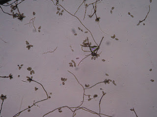
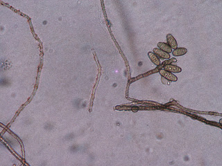
Next, I observed my plate taken from the shower (Fig. 4). I also observed this fungus under the microscope, but was unable to make an ID (Figs. 5-7). This particular fungus exhibited clusters of pill-shaped spores that exhibited at most 4 divisions.
 |
| Fig. 4 Plate taken from shower |
 |
| Fig. 5 Conidiophores of shower fungus |
 |
| Fig. 6 Condiophores and spores of shower fungus |
 |
| Fig. 7 Conidiophores and spores of shower fungus |
 |
| Fig. 8 Light green fungus isolated from pigs |
 |
| Fig. 9 Conidiophore at 40x magnification |
 |
| Fig. 10 Hypha showing septa |
 |
| Fig. 11 Observation of entire plate under the compound microscope |
 |
| Fig. 12 Observation of entire plate under the compound microscope |
 |
| Fig. 13 Utilization of a tape mount in order to better observe structures |
 |
| Fig. 14 Moldy cricket found in a ditch |
 |
| Fig. 15 Abandoned wasp nest |
Looks like I need to spend an ample amount of time trying to figure out all of my samples!
All for now,
C
No comments:
Post a Comment