Collection: Longview, TX
My parents live in the Piney Woods of East Texas, where it is very humid and there are always neat fungi growing everywhere. So I took the opportunity to collect some samples when I visited them this semester.
The first collection I made was from a pile of Sweetgum logs that have been sitting in one of our fields for over a year (Fig. 1). There were obvious colonies of fungi on these pieces of wood, particularly those in the middle of the pile (Figs. 2, 3). I utilized the same sterile swab technique that I have used previously to take samples.
 |
| Fig. 1 Sweetgum logs in one of our fields |
 |
| Fig. 2 Obvious fungal colonies on Sweetgum logs |
 |
| Fig. 3 Obvious fungal colonies on Sweetgum logs |
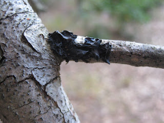
The second collection I made while in Longview was off of a living tree in our woods. This was a large black fungus covering the external surface of the trunk and the branches of the tree (Figs. 4, 5). In order to collect this fungus, I cut off the entire fungus with a scalpel that I had previously sterilized and placed it inside of a brown paper bag for transport to the lab.
 |
| Fig. 4 Black fungus growing on external surface of tree trunk |
 |
| Fig. 5 Black fungus growing on external surface of tree branch |
 |
| Fig. 6 Black and white mold growing at the base of the shower. |
Upon arriving back at the lab, I plated everything onto 1/2 strength PDA except for the shower samples, which I plated on water agar. I figured that if this mold can grow on shower tiles, then it could grow on water agar. For the whole chunks of fungus that I cut from the tree bark, I placed one whole body onto the agar to see what I could grow.
After 4 days, all of my plates showed extensive growth (Figs. 7-9). The fungal body that I plated directly on the 1/2 strength PDA showed at least 2 different colonies (at least macroscopically they looked different) (Fig. 7). The plate containing fungus taken from the shower showed some particularly interesting (and nasty) growth (Fig. 8). This particular plate exhibited very dark, hard structures, most of which looked like pycnidia (I believe). When slide-mounted, these structures would burst and release a billion tiny spores (Fig. 9). These structures were also entangled in an extensive amount of hyphae (Fig. 10). I did not try to key either of these fungi out at this time.
 |
| Fig. 7 Plate showing entire fungal body (black structure in the middle), with at least two different colonies |
 |
| Fig. 8 Colonies growing from shower sample |
 |
| Fig. 9 Putative pycnidia that have ruptured and released many tiny spores |
 |
| Fig. 10 Entanglement of hyphae surrounding pycnidia |
Conclusion
Looks like I am making at least a little bit of progress with the unknowns. At least they aren't all Rhizopus or Mucor!
All for now,
C
No comments:
Post a Comment