For the first actual lab, I learned many new techniques used for viewing and identifying morphological structures of fungi. These techniques include slide mounting with both squash mount and tape mounts, cell-counting with a hemacytometer, and utilizing a dichotomous key for identification of fungi. Several cultures of various fungal genera were also available for observation and identification. Below, I will summarize my experience with each technique as well as provide information regarding the different fungal cultures I observed.
Techniques.
Squash-mount.
The first technique I learned was the squash mount. A drop of sterile water is first placed in the middle of a clean glass slide. While keeping the culture plate at the base of the bunsen burner, a probe is then sterilized via open flame, the lid of the plate is opened slightly, and the probe is cooled by sticking the tip in a clean area of agar. Fungal material is then gently scraped from the culture and the lid of the plate is closed again. The material that has adhered to the probe can then be deposited into the water droplet on the slide. After this transfer, the coverslip is then placed on top of the inoculated droplet and air bubbles are pressed out from under the slip with the blunt end of the probe. Now the slide is ready for viewing under the compound microscope. Hooray! I made approximately 20 different slides with this technique.
Tape-mount.
Another slide-making technique that I had never heard of prior to last week is called a tape mount. Again, a drop of sterile water is placed in the center of a clean glass slide (the water is optional, but I found that it helps with the quality of the image produced by the microscope). Instead of inoculating with a sterile tool, a length of scotch tape (approximately 2cm) is taken and is just barely allowed it to make contact with the fungal substrate. This is then placed over the drop of water on the slide. Ideally, the ends of the tape are supposed to stay dry and adhere to the slide. This type of mount is supposed to be good for seeing conidiophores; however, I found out very quickly that I am not very good at making these types of mounts as I kept collecting a ridiculous amount of spores. This made it difficult to see anything under the microscope. I made around 5 slides this way and decided that I was going to have more luck using the first technique. However, I will practice this technique every lab period.
Cell counting.
We watched a video on the methodology behind counting cells using a hemocytometer (http://www.youtube.com/watch?v=pP0xERLUhyc). A dilution is made of the appropriate fungus and squirted into a special (very expensive) glass slide with tiny quadrants. These quadrants (and the use of a hand counter) allow for simple counting of both viable and dead cells. I practiced making one dilution and looked at it under the microscope; however, I did not actually try to count any cells. I definitely need more practice on this and hope to gain more experience as the labs continue.
Dichotomous key
We were introduced to several keys that will be helpful in identifying fungus this semester. The one I will likely be utilizing the most is the Illustrated Genera of Imperfect Fungi by Barnett and Hunter. I have already purchased a copy and am excited to start using it in the next lab. Although I did look through the pictures in the key to compare with what I was seeing on my slides, I did not actually start going through the couplets. I would like to think that I am wise enough now to know that it is more time-efficient and less stressful to get acquainted with a new type of organism by a few casual observations before trying to delve into details that I am not familiar, or comfortable, with identifying just yet. As Lab 1 was the very first time I had ever observed a fungus under the scope, I was not about to try to take it through a key. I will begin keying out specimens this week in lab and hopefully become proficient at it by the end of the semester.
Just so it doesn't seem like I am a TOTAL noob, I do have a lot of experience working with both compound and dissecting scopes. I have made thousands of slides of nearly microscopic mites and extensively used dichotomous keys to ID numerous types of arthropods. So, even though I have never used these particular techniques before doesn't mean that they are completely foreign to me.
Observations of Fungal Cultures
We were allowed to observe different fungal cultures in this lab in order to start getting familiar with basic morphology of different genera. I observed the followings fungi:
1. Aspergillua niger
2. Alternaria brassicicola
3. Thielaviopsis basicola
4. Cladosporium sp.
Fungus 1: Aspergillus niger.
I was able to see many conidia, but it was very hard to find the conidiophore of this fungus. I spent a lot of time doing different types of mounts, but I never saw one on my own. The conidia were spherical and formed long chains (Fig. 1).
 |
| Fig. 1 Hand-drawing of the spherical conidia of Aspergillus niger from my lab notebook. |
Fungus 2: Alternaria brassicicola.
I was still only able to see the conidia on my own slides, but I believe I saw the conidiophore of these on some of my labmate's slides. Conidia were linked in short and long chains, had a more rectangular appearance, and usually terminated in a knob-like structure (Fig. 2).
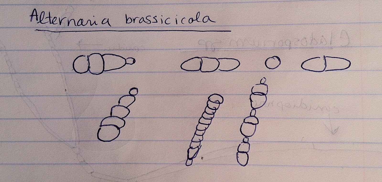 |
| Fig. 2 Hand-drawing of the conidia of Alternaria brassicocola from my lab notebook. |
Fungus 3. Thielaviopsis bassicola.
I finally saw a conidiophore with this species! I was very excited about this. Conidia occurred in what seemed like two different types of spores. The first type consisted of very long, thin, rectangular cells, and the second type consisted of more oval-like spores. I believe the second type is referred to as the aleuriospore, in which the spore is further subdivided by a variable number of cells (Fig. 3).
 |
| Fig. 3 Hand-drawing of conidia and conidiophore of Thielaviopsis bassicola from my lab notebook. |
Fungus 4. Cladosporium sp.
I was also able to easily see the conidiophore on this fungus. It was very long, slender, and rectangular, and terminated in many small spherical conidia (Fig. 4).
 |
| Fig. 4 Hand-drawing conidiophore and conidia of Cladosporium sp. from my lab notebook. |
Conclusion
Overall, I learned many new techniques for visualizing and identifying fungi in this first laboratory. However, it will definitely take time to get used to looking at these structures and placing a name to them. I look forward to honing my morphological identification skills in this class.
-C
No comments:
Post a Comment